What Are The Signals Of Calcaneal Spur

Overview
Heel Spurs should be called a traction spurs because they grow in the same direction that the tendons pull away from the heel bone, which is why it can occur on the bottom of the heel (Plantar Fasciitis) and on the back of the heel (Achilles Tendonitis). Some patients may only develop one type of heel spur, but both these problems are closely related so it's not unusual for a patient to have both heel spurs. It's important to note though that most heel spurs aren't the cause of your heel pain.
Causes
One of the most common causes for the development of heel spurs is the wearing of shoes that are too tight. That?s why more women suffer from heel spurs more than men. Athletes who tend to stress their feet a lot, people are overweight who have more pressure on their lower extremities and the elderly also tend to suffer more from heel spurs.
Symptoms
Symptoms may be similar to those of plantar fasciitis and include pain and tenderness at the base of the heel, pain on weight bearing and in severe cases difficulty walking. The main diagnosis of a heel spur is made by X-ray where a bony growth on the heel can be seen. A heel spur can occur without any symptoms at all and the athlete would never know they have the bony growth on the heel. Likewise, Plantar fasciitis can occur without the bone growth present.
Diagnosis
A thorough medical history and physical exam by a physician is always necessary for the proper diagnosis of heel spurs and other foot conditions. X rays of the heel area are helpful, as excess bone production will be visible.
Non Surgical Treatment
Heel spurs are considered a self-limited condition, which means that by making small alterations in your lifestyle and regular routines you can often control the condition. The goal is to relieve pain, reduce friction and transfer pressure from your sensitive foot areas. By eliminating the cause of the heel spur and plantar fasciitis (i.e. better shoes, orthotics to fix your gait, losing weight) will help reduce the pressure put on your fascia and heel and can reduce the inflammation caused by your heel spur. Failure to see improvements after conservative treatments may make surgery your only option.
Surgical Treatment
Surgery to correct for heel spur syndrome is a common procedure which releases plantar fascia partially from its attachment to the calcaneous (heel bone). This part of the surgery is called a plantar fasciotomy due to the fact the fascia is cut. This is most often done through an open procedure as any heel spur or bursa can be removed at the same time. If the spur is not removed during the surgery, it will probably be just as successful, as the large spur is not the true problem. Some physicians use an endoscopic approach (EPF) where a small camera aids the physician during surgery with typically smaller incisions on each side of your foot.
Prevention
To prevent this condition, wearing properly fitted shoes with good arch support is very important. If a person is overweight, weight loss can help diminish stress on the feet and help prevent foot problems. For those who exercise frequently and intensely, proper stretching is always necessary, especially when there is an increase in activities or a change in running technique. It is not recommended to attempt to work through the pain, as this can change a mild case of heel spurs and plantar fascitis into a long-lasting and painful episode of the condition.
Bursitis Of The Foot Bursal Cyst
Overview
Retrocalcaneal bursitis is sometimes difficult to differentiate from Achilles tendinitis, at least symptomatically. Both are most uncomfortable during the push-off phase of gait, are most severely painful in the morning and with walking after sitting for a period of time, and generally worsen with activity. Most practitioners make the distinction between the two simply on the basis of location of pain and tenderness. Generally, Achilles tendinitis is felt an inch or two higher than this form of bursitis.
Causes
Bursitis can develop for several reasons, including repetitively engaging in the same motion, or example, lifting objects above your head for work. Putting a lot of pressure on a bursa for an extended period of time. Leaning on your elbows or kneeling (for example, to lay carpet) can cause bursitis in the elbows or knees. If you sit for long periods of time, especially on hard surfaces, you may develop bursitis in your hip. Wearing shoes with a stiff back that rubs against the back of the ankle can cause Achilles tendon bursitis. Trauma. The bursae at the knee and elbow are close to the surface of the skin, and if you fall directly on your elbow or the knee, you can rupture, injure or puncture a bursa. Infection. Known as septic bursitis, it?s the result of bacteria infecting a bursa. It can occur from an infection traveling from another site or following an accident that ruptures the bursa. Even scraping the skin on your elbow or getting a mosquito bite that breaks the skin near the olecranon bursa (near the elbow) can lead to bursitis. Other joint disorders, such as rheumatoid arthritis, osteoarthritis and gout, or health conditions.
Symptoms
Unlike Achilles tendinitis, which tends to manifest itself slightly higher on the lower leg, Achilles tendon bursitis usually creates pain and irritation at the back of the heel. Possible signs of bursitis of the Achilles tendon include difficulty to rise on toes. Standing on your toes or wearing high heels may increase the heel pain. Inflammation and tenderness. The skin around your heel can become swollen and warm to the touch. Redness may be visible. Pain in the heel. Pain tends to become more prominent when walking, running, or touching the inflamed area. Stiffness. The back of your ankle may feel a little stiff due to the swelling of the bursa.
Diagnosis
The diagnosis is based on the symptoms and an examination. For anterior Achilles tendon bursitis, doctors use x-rays to rule out a fracture of the heel bone or damage to the heel bone caused by rheumatoid arthritis or other inflammatory arthritis.
Non Surgical Treatment
The patient with retrocalcaneal bursitis should be instructed to apply ice to the posterior heel and ankle in the acute period of the bursitis. Icing can be performed several times a day, for 15-20 minutes each. Some clinicians also advocate the use of contrast baths. Gradual progressive stretching of the Achilles tendon may help relieve impingement on the subtendinous bursa and can be performed in the following manner. Stand in front of a wall, with the affected foot flat on the floor. Lean forward toward the wall until a gentle stretching is felt within the ipsilateral Achilles tendon. Maintain the stretch for 20-60 seconds and then relax. Perform the stretches with the knee extended and then again with the knee flexed. To maximize the benefit of the stretching program, repeat the above steps for several stretches per set, several times daily. Avoid ballistic (ie, abrupt, jerking) stretches. Other treatment options are microcurrent therapy and corticosteriod injection into the retrocalcaneal bursa. If conservation treatment fails then surgery is indicated.
Surgical Treatment
Only if non-surgical attempts at treatment fail, will it make sense to consider surgery. Surgery for retrocalcanel bursitis can include many different procedures. Some of these include removal of the bursa, removing any excess bone at the back of the heel (calcaneal exostectomy), and occasionally detachment and re-attachment of the Achilles tendon. If the foot structure and shape of the heel bone is a primary cause of the bursitis, surgery to re-align the heel bone (calcaneal osteotomy) may be considered. Regardless of which exact surgery is planned, the goal is always to decrease pain and correct the deformity. The idea is to get you back to the activities that you really enjoy. Your foot and ankle surgeon will determine the exact surgical procedure that is most likely to correct the problem in your case. But if you have to have surgery, you can work together to develop a plan that will help assure success.
Hammertoes Treatment Options
 Overview
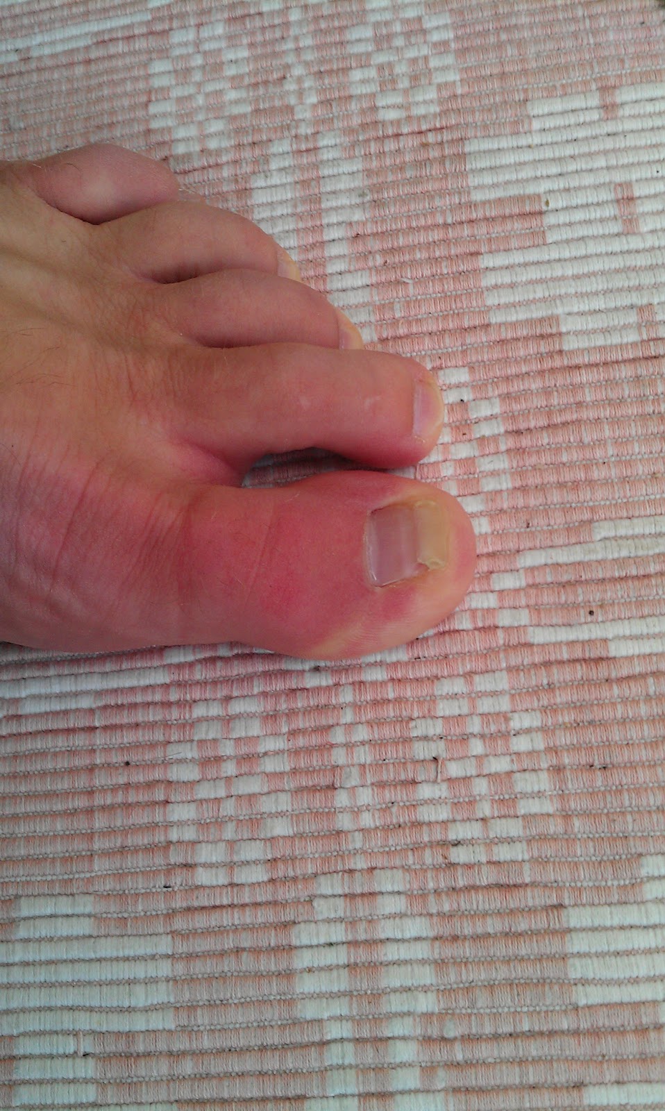
A hammertoes is a toe that is bent because of a weakened muscle. The weakened muscle makes the tendons (tissues that connect muscles to bone) shorter, causing the toes to curl under the feet. Hammertoes can run in families. They can also be caused by shoes that are too short. Hammertoes can cause problems with walking and can lead to other foot problems, such as blisters, calluses, and sores. Splinting and corrective footwear can help in treating hammertoes. In severe cases, surgery to straighten the toe may be necessary.
Causes
Your toe contains two joints that allow it to bend at the middle and bottom. A hammertoe occurs when the middle joint becomes dislocated. Common causes of this hammertoes joint dislocation include a toe injury, arthritis, a high foot arch, wearing shoes that don?t fit properly, tightened ligaments or tendons in the foot, pressure from a bunion (when your big toe points inward toward your second toe) Spinal cord or peripheral nerve damage may cause all of your toes to curl downward.
 Symptoms
A hammertoe may be present but not always painful unless irritated by shoes. One may have enlarged toe joints with some thickened skin and no redness or swelling. However, if shoes create pressure on the joint, the pain will usually range from pinching and squeezing to sharp and burning. In long standing conditions, the dislocated joints can cause the pain of arthritis.
Diagnosis
Your doctor is very likely to be able to diagnose your hammertoe simply by examining your foot. Even before that, he or she will probably ask about your family and personal medical history and evaluate your gait as you walk and the types of shoes you wear. You'll be asked about your symptoms, when they started and when they occur. You may also be asked to flex your toe so that your doctor can get an idea of your range of motion. He or she may order x-rays in order to better define your deformity.
Non Surgical Treatment
Conservative treatment is the first choice, often starting with a change of shoes to ones that have soft, larger toe spaces. Toe exercises may be prescribed to stretch and strengthen the toe muscles. Over-the-counter straps, cushions or non-medicated corn pads may be recommended to help relieve your symptoms.
Surgical Treatment
Hammer toe can be corrected by surgery if conservative measures fail. Usually, surgery is done on an outpatient basis with a local anesthetic. The actual procedure will depend on the type and extent of the deformity. After the surgery, there may be some stiffness, swelling and redness and the toe may be slightly longer or shorter than before. You will be able to walk, but should not plan any long hikes while the toe heals, and should keep your foot elevated as much as possible.
Hammertoes Treatment At Home
 Overview
hammertoes (hammertoe) is a deformity of the second, third, or fourth toes. In this condition, the toe is bent at the middle joint, into an upward position, causing it to resemble a hammer (sometimes decribed as ?curled toes?). Left untreated, hammer toes can become inflexible and require surgery. Toes which take on a curled appearance are hammer toes. Mallet toe is a similar condition, but affects the upper joint of a toe.
Causes
Shoes that narrow toward the toe force the smaller toes into a bent upward position. This makes the toes rub against the inside of the shoe, and creates corns and calluses, aggravating the toes further. If the shoes have a high heel, the feet are forced forward and down, squeezing the toes against the front of the shoe, which increases the pressure on the toes and makes them bend further. Eventually, the toe muscles become unable to straighten the toe.
 Symptoms
If the toes remain in the hammertoe position for long periods, the tendons on the top of the foot will tighten over time because they are not stretched to their full length. Eventually, the tendons shorten enough that the toe stays bent, even when shoes are not being worn. The symptoms Hammer toes of hammertoe include a curling toe, pain or discomfort in the toes and ball of the foot or the front of the leg, especially when toes are stretched downward, thickening of the skin above or below the affected toe with the formation of corns or calluses, difficulty finding shoes that fit well. In its early stages, hammertoe is not obvious. Frequently, hammertoe does not cause any symptoms except for the claw-like toe shape.
Diagnosis
The treatment options vary with the type and severity of each hammer toe, although identifying the deformity early in its development is important to avoid surgery. Your podiatric physician will examine and X-ray the affected area and recommend a treatment plan specific to your condition.
Non Surgical Treatment
Conservative treatment is limited to accommodation, not correction, of the deformity, though some patients find the relief they can get from these options to be more than enough to put off or even avoid surgery. These include better Footwear. Shoe gear with a wider toe box and higher volume causes less friction to the toes. Toe Braces and Strapping. Some toe braces and strapping techniques take some pressure off the toes during gait. Custom molded orthotics can redistribute the forces through the tendons that control the toe, lessening the pain and extent of the deformity.The calluses on the toe and the ball of the foot can be shaved occasionally to reduce some pain and pressure, although they will return due to the constant deformity.
Surgical Treatment
If these treatments are not sufficient at correcting the hammer toe, an operation to straighten the toe may be necessary. This is often performed in conjunction with surgery for a bunion deformity. The surgical treatment of a hammer toe can consist of either cutting the tendons to relieve the pressure that causes the deformity, or fusing the toe so that it points straight permanently.
What Causes Over-Pronation
Overview
Over Pronation (Flat Feet) refers to the biomechanical shock-absorbing motion of the ankle, foot and lower leg. It is the natural inward flexing motion of the lower leg and ankle. Standing, walking, and running cause the ankle joint to pronate which in turn helps the body to absorb shock and allows it to control balance. An ankle joint that is too flexible causes more pronation than desired. This common condition is called Over- Pronation (sometimes referred to as "Flat Feet"). This foot condition places an extreme degree of strain on various connective tissues of the ankle, foot, and knee. If this condition is not addressed foot pain and toe deformities such as bunions and hammer toes (just to name a couple) may develop. Hip and lower back pain may also be residual results from this condition.
Causes
There is a relationship between biomechanics and injury that is specific to each body part. Overall though, poor mechanics will either increase the landing forces acting on the body or increase the work to be done by the muscles. Both increase the stress, which, depending on the individual and the amount of running can become excessive and cause injury.
Symptoms
Because pronation is a twisting of the foot, all of the muscles and tendons which run from the leg and ankle into the foot will be twisted. In over-pronation, resulting laxity of the soft tissue structures of the foot and loosened joints cause the bones of the feet shift. When this occurs, the muscles which attach to these bones must also shift, or twist, in order to attach to these bones. The strongest and most important muscles that attach to our foot bones come from our lower leg. So, as these muscles course down the leg and across the ankle, they must twist to maintain their proper attachments in the foot. Injuries due to poor biomechanics and twisting of these muscles due to over-pronation include: shin splints, Achilles Tendonitis, generalized tendonitis, fatigue, muscle aches and pains, cramps, ankle sprains, and loss of muscular efficiency (reducing walking and running speed and endurance). Foot problems due to over-pronation include: bunions, heel spurs, plantar fasciitis, fallen and painful arches, hammer toes, and calluses.
Diagnosis
You can test for pronation by looking at the leg and foot from the back. Normally you can see the Achilles Tendon run straight down the leg into the heel. If the foot is pronated, the tendon will run straight down the leg, but when it lies on the heel it will twist outward. This makes the inner ankle bone much more prominent than the outer ankle bone.
Non Surgical Treatment
Over-pronation and the problems that go with it are treated with shoe inserts called arch supports or orthotics. You can buy orthotics at a pharmacy or athletic shoe store or they can be custom made. Make sure the arch supports are firm. If you can easily bend them in half, they may be too flexible.
Surgical Treatment
Subtalar Arthroereisis. The ankle and hindfoot bones/midfoot bones around the joint are fused, locking the bones in place and preventing all joint motion. This may also be done in combination with fusion at other joints. This is a very aggressive option usually reserved for extreme cases where no joint flexibility is present and/or the patient has severe arthritic changes in the joint.
What Exactly Is Severs Disease?
Overview
Sever?s Disease (calcaneal aphophysitis) is not really a disease, but more a repetitious strain injury. This is the most common cause of kids heel pain seen at Podiatry Care. Active children in football, soccer, basketball, netball and tennis with this type of foot pain complain of pain in the region of the heel bone particularly after exercise. In severe cases, children will complain of pain during exercise as well. It is a frequent cause of heel pain in children, particularly in the very active child. It is most often seen in children between the ages of 8 to 15 years as the growth plate is not fully developed or calcified at this time.
Causes
Your child is most at risk for this condition when he or she is in the early part of the growth spurt in early puberty. Sever's disease is most common in physically active girls 8 years to 10 years of age and in physically active boys 10 years to 12 years of age. Soccer players and gymnasts often get Sever's disease, but children who do any running or jumping activity may also be at an increased risk. Sever's disease rarely occurs in older teenagers because the back of the heel has typically finished growing by 15 years of age.
Symptoms
The most prominent symptom of Sever's disease is heel pain which is usually aggravated by physical activity such as walking, running or jumping. The pain is localized to the posterior and plantar side of the heel over the calcaneal apophysis. Sometimes, the pain may be so severe that it may cause limping and interfere with physical performance in sports. External appearance of the heel is almost always normal, and signs of local disease such as edema, erythema (redness) is absent. The main diagnostic tool is pain on medial- lateral compression of the calcaneus in the area of growth plate, so called squeeze test. Foot radiographs are usually normal. Therefore the diagnosis of Sever's disease is primarily clinical.
Diagnosis
Sever?s disease can be diagnosed based on your history and symptoms. Clinically, your physiotherapist will perform a "squeeze test" and some other tests to confirm the diagnosis. Some children suffer Sever?s disease even though they do less exercise than other. This indicates that it is not just training volume that is at play. Foot and leg biomechanics are a predisposing factor. The main factors thought to predispose a child to Sever?s disease include a decrease in ankle dorsiflexion, abnormal hind foot motion eg overpronation or supination, tight calf muscles, excessive weight-bearing activities eg running.
Non Surgical Treatment
Treatment may consist of one or more of the following, Elevating the heel, Stretching hamstring and calf muscles 2-3 times daily, Using R.I.C.E. (Rest, Ice, Compression, Elevation), Foot orthotics, Medication, Physical therapy, Icing daily (morning), Heating therapy, Open back shoe are best and avoid high heel shoe. The Strickland Protocol has shown a positive response in patients with a mean return to sport in less than 3 weeks.
Prevention
As with all overuse injuries, it is important to warm up sufficiently before you exercise and warm down afterwards. You should build up any alterations in the intensity of your training gradually, and never continue exercising with weakened or fatigued muscles. Replace any worn or tattered shoes, as in this condition they become useless for absorbing shock and protecting the feet.
Collapsed Arch Pain Relief
Overview
Posterior tibial tendon dysfunction is one of several terms to describe a painful, progressive flatfoot deformity in adults. Other terms include posterior tibial tendon insufficiency and adult acquired flatfoot. The term adult acquired flatfoot is more appropriate because it allows a broader recognition of causative factors, not only limited to the posterior tibial tendon, an event where the posterior tibial tendon looses strength and function. The adult acquired flatfoot is a progressive, symptomatic (painful) deformity resulting from gradual stretch (attenuation) of the tibialis posterior tendon as well as the ligaments that support the arch of the foot.

Causes
Plantar Fasciitis is commonly the cause of most arch pain. The bands of fibrous tissue in the arches of the feet become inflamed. Plantar Fasciitis is associated with early morning arch pain, from the plantar fascia tightening and contracting during the night when there is no strain on the bands. Arch pain occurs when there are extended periods of standing or walking, resulting in prolonged tension on the plantar fascia which in turn causes inflammation and irritation. While plantar fasciitis normally affects middle aged men and women, younger athletes are affected by arch pain because of the repetitive movement of certain sports, which causes damage to the fibrous tissue.
Symptoms
Many people have no symptoms, and the condition is discovered only by chance when an X-ray of the foot is obtained for some other problem. When symptoms occur, there is usually foot pain that begins at the outside rear of the foot. The pain tends to spread upward to the outer ankle and to the outside portion of the lower leg. Symptoms usually start during a child's teenage years and are aggravated by playing sports or walking on uneven ground. In some cases, the condition is discovered when a child is evaluated for unusually frequent ankle sprains.
Diagnosis
In more difficult cases of plantar fasciitis you should see your foot health professional for a thorough examination. They will find out why your arch or heel pain occurred in the first place and devise a treatment plan to relieve your pain and prevent it from reoccurring. They will evaluate your feet, walking pattern (gait), shoes, activities, exercise methods, and other relevant information and then devise your treatment plan.
Non Surgical Treatment
Cortisone, a type of steroid, is a powerful anti-inflammatory medication. It can be injected into the plantar fascia to reduce inflammation and pain. Your doctor may limit your injections. Multiple steroid injections can cause the plantar fascia to rupture (tear), which can lead to a flat foot and chronic pain. Supportive shoes and orthotics. Shoes with thick soles and extra cushioning can reduce pain with standing and walking. As you step and your heel strikes the ground, a significant amount of tension is placed on the fascia, which causes microtrauma (tiny tears in the tissue). A cushioned shoe or insert reduces this tension and the microtrauma that occurs with every step. Soft silicone heel pads are inexpensive and work by elevating and cushioning your heel. Pre-made or custom orthotics (shoe inserts) are also helpful. Most people sleep with their feet pointed down. This relaxes the plantar fascia and is one of the reasons for morning heel pain. A night splint stretches the plantar fascia while you sleep. Although it can be difficult to sleep with, a night splint is very effective and does not have to be used once the pain is gone. Your doctor may suggest that you work with a physical therapist on an exercise program that focuses on stretching your calf muscles and plantar fascia. In addition to exercises like the ones mentioned above, a physical therapy program may involve specialized ice treatments, massage, and medication to decrease inflammation around the plantar fascia. Extracorporeal shockwave therapy (ESWT). During this procedure, high-energy shockwave impulses stimulate the healing process in damaged plantar fascia tissue. ESWT has not shown consistent results and, therefore, is not commonly performed. ESWT is noninvasive-it does not require a surgical incision. Because of the minimal risk involved, ESWT is sometimes tried before surgery is considered.

Surgical Treatment
The soft tissue surgeries usually would include a lengthening of the Achilles tendon, releasing of the plantar fascia as well as tendon transfers. These procedures are usually done in conjunction with bony procedures such as calcaneal osteotomies (to lower the heel bone and get it more under the leg itself), as well as metatarsal osteotomies. These procedures usually involve either cutting or fusion of the bones, and placement of fixation devices to allow the bones to heal. Healing time is usually at least 6-8 weeks and usually the patient must be non-weight bearing during the healing process. These types of surgical corrections are usually reserved for the more difficult, painful and deformed feet. They can require more surgeries down the line. These procedures are usually the last resort after all other modes of treatment have been exhausted (except in children where it is usually best to treat the deformity early). There are many different degrees of high arched feet and these procedures should be left for the more extreme cases. These cases usually require a very high degree of surgical skill and should only be done by those who frequently perform these types of cases.
Prevention
Maintain a healthy weight, Use insoles to support your arches, Limit how often you wear high heels, Use proper shoes, especially when exercising to evenly distribute weight through your foot.
Stretching Exercises
Easy Beginner Version. Start with your bare foot on a flat surface, toes spread out. Place a penny under the ball of your foot and the end of a pen under the middle of your arch (sticking out from the inside of your foot). Activate your arch by flexing your arch muscle. You should feel the muscles on the ball of your foot pushing down on the penny, but your arch shouldn't be pushing down on the pen. These tools help you (1) avoid rolling your foot and (2) avoid pressing down with your toes (as an extra tip, you can slide a business card under your toes before doing the exercise-when you activate your arch, you should be able to slide the business card out easily with your fingers). Do your best to keep your toes relaxed. Advanced Version. Once you're ready to move on, you can try this advanced version. It builds on the above exercise to incorporate full body twisting and balance, helping you to maintain proper arches while you move. Using the same ideas from above, stand on a flat surface in your bare feet with a penny under the ball of your foot and the end of a pen under your arch. This time, stand with your back a few inches away form a wall or a door. Lift your other leg (the one without the penny or pen) and stand on one foot. Use the wall for balance, if necessary. Lift one arm and stretch it across your body until you touch the wall or door on the opposite side, maintaining a straight back. Keep your foot straight and your arch on the penny but above the pen. Your arch will want to follow the movement and roll off, but you will need to activate it to stay stable during the movement. Lift your other arm and stretch it across the opposite side of your body, still keeping your arch in place.
Posterior tibial tendon dysfunction is one of several terms to describe a painful, progressive flatfoot deformity in adults. Other terms include posterior tibial tendon insufficiency and adult acquired flatfoot. The term adult acquired flatfoot is more appropriate because it allows a broader recognition of causative factors, not only limited to the posterior tibial tendon, an event where the posterior tibial tendon looses strength and function. The adult acquired flatfoot is a progressive, symptomatic (painful) deformity resulting from gradual stretch (attenuation) of the tibialis posterior tendon as well as the ligaments that support the arch of the foot.
Causes
Plantar Fasciitis is commonly the cause of most arch pain. The bands of fibrous tissue in the arches of the feet become inflamed. Plantar Fasciitis is associated with early morning arch pain, from the plantar fascia tightening and contracting during the night when there is no strain on the bands. Arch pain occurs when there are extended periods of standing or walking, resulting in prolonged tension on the plantar fascia which in turn causes inflammation and irritation. While plantar fasciitis normally affects middle aged men and women, younger athletes are affected by arch pain because of the repetitive movement of certain sports, which causes damage to the fibrous tissue.
Symptoms
Many people have no symptoms, and the condition is discovered only by chance when an X-ray of the foot is obtained for some other problem. When symptoms occur, there is usually foot pain that begins at the outside rear of the foot. The pain tends to spread upward to the outer ankle and to the outside portion of the lower leg. Symptoms usually start during a child's teenage years and are aggravated by playing sports or walking on uneven ground. In some cases, the condition is discovered when a child is evaluated for unusually frequent ankle sprains.
Diagnosis
In more difficult cases of plantar fasciitis you should see your foot health professional for a thorough examination. They will find out why your arch or heel pain occurred in the first place and devise a treatment plan to relieve your pain and prevent it from reoccurring. They will evaluate your feet, walking pattern (gait), shoes, activities, exercise methods, and other relevant information and then devise your treatment plan.
Non Surgical Treatment
Cortisone, a type of steroid, is a powerful anti-inflammatory medication. It can be injected into the plantar fascia to reduce inflammation and pain. Your doctor may limit your injections. Multiple steroid injections can cause the plantar fascia to rupture (tear), which can lead to a flat foot and chronic pain. Supportive shoes and orthotics. Shoes with thick soles and extra cushioning can reduce pain with standing and walking. As you step and your heel strikes the ground, a significant amount of tension is placed on the fascia, which causes microtrauma (tiny tears in the tissue). A cushioned shoe or insert reduces this tension and the microtrauma that occurs with every step. Soft silicone heel pads are inexpensive and work by elevating and cushioning your heel. Pre-made or custom orthotics (shoe inserts) are also helpful. Most people sleep with their feet pointed down. This relaxes the plantar fascia and is one of the reasons for morning heel pain. A night splint stretches the plantar fascia while you sleep. Although it can be difficult to sleep with, a night splint is very effective and does not have to be used once the pain is gone. Your doctor may suggest that you work with a physical therapist on an exercise program that focuses on stretching your calf muscles and plantar fascia. In addition to exercises like the ones mentioned above, a physical therapy program may involve specialized ice treatments, massage, and medication to decrease inflammation around the plantar fascia. Extracorporeal shockwave therapy (ESWT). During this procedure, high-energy shockwave impulses stimulate the healing process in damaged plantar fascia tissue. ESWT has not shown consistent results and, therefore, is not commonly performed. ESWT is noninvasive-it does not require a surgical incision. Because of the minimal risk involved, ESWT is sometimes tried before surgery is considered.
Surgical Treatment
The soft tissue surgeries usually would include a lengthening of the Achilles tendon, releasing of the plantar fascia as well as tendon transfers. These procedures are usually done in conjunction with bony procedures such as calcaneal osteotomies (to lower the heel bone and get it more under the leg itself), as well as metatarsal osteotomies. These procedures usually involve either cutting or fusion of the bones, and placement of fixation devices to allow the bones to heal. Healing time is usually at least 6-8 weeks and usually the patient must be non-weight bearing during the healing process. These types of surgical corrections are usually reserved for the more difficult, painful and deformed feet. They can require more surgeries down the line. These procedures are usually the last resort after all other modes of treatment have been exhausted (except in children where it is usually best to treat the deformity early). There are many different degrees of high arched feet and these procedures should be left for the more extreme cases. These cases usually require a very high degree of surgical skill and should only be done by those who frequently perform these types of cases.
Prevention
Maintain a healthy weight, Use insoles to support your arches, Limit how often you wear high heels, Use proper shoes, especially when exercising to evenly distribute weight through your foot.
Stretching Exercises
Easy Beginner Version. Start with your bare foot on a flat surface, toes spread out. Place a penny under the ball of your foot and the end of a pen under the middle of your arch (sticking out from the inside of your foot). Activate your arch by flexing your arch muscle. You should feel the muscles on the ball of your foot pushing down on the penny, but your arch shouldn't be pushing down on the pen. These tools help you (1) avoid rolling your foot and (2) avoid pressing down with your toes (as an extra tip, you can slide a business card under your toes before doing the exercise-when you activate your arch, you should be able to slide the business card out easily with your fingers). Do your best to keep your toes relaxed. Advanced Version. Once you're ready to move on, you can try this advanced version. It builds on the above exercise to incorporate full body twisting and balance, helping you to maintain proper arches while you move. Using the same ideas from above, stand on a flat surface in your bare feet with a penny under the ball of your foot and the end of a pen under your arch. This time, stand with your back a few inches away form a wall or a door. Lift your other leg (the one without the penny or pen) and stand on one foot. Use the wall for balance, if necessary. Lift one arm and stretch it across your body until you touch the wall or door on the opposite side, maintaining a straight back. Keep your foot straight and your arch on the penny but above the pen. Your arch will want to follow the movement and roll off, but you will need to activate it to stay stable during the movement. Lift your other arm and stretch it across the opposite side of your body, still keeping your arch in place.